


The first combination of 3D printing technology and medicine was the replacement of the lower collar bone for patients with osteomyelitis, achieving satisfactory results. At present, 3D printing technology is most widely used in the field of orthopedics. With the increasing awareness of 3D printing technology among medical workers, it has gradually been applied in other systems of the human body. The application of 3D printing technology in the implementation of treatment plans has also achieved remarkable results, with representative enterprises in this area such as Senna Digital Medical.
1、 Hepatobiliary system
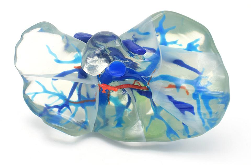
Due to the presence of portal vein, hepatic vein, and hepatic artery in the liver, and the inferior vena cava passing through the liver, how to completely remove the tumor, preserve normal liver tissue to the maximum extent, and reduce intraoperative and postoperative complications has always been one of the challenges in liver surgery. Hepatobiliary surgery experts have applied 3D printing technology to the resection of liver tumors. 3D printed models can display the spatial relationship between liver tumors and intrahepatic vessels in a three-dimensional manner, helping to define the pre resection surface of the liver and ensuring accurate implementation of actual surgery. 3D printing technology has achieved a leapfrog transformation from 3D images to three-dimensional physical models. When performing liver tumor resection, 3D printing technology helps with preoperative safety assessment, key anatomical site localization, real-time navigation surgery, and anatomical liver resection. By adjusting the 3D printed model at any time during surgery and placing it in the optimal anatomical position for comparison with real-time surgery, it can provide intuitive real-time navigation for key surgical steps. By accurately locating the lesion and determining the surgical resection plane, real-time guidance is provided for the separation of important vessels and the resection of tumor lesions, achieving complete resection of the lesion and avoiding secondary damage to important anatomical structures, thereby improving the curative effect of surgery and reducing surgical risk, while reducing surgical time and complications.
2、 Urinary system
In the clinical treatment of kidney tumors, 3D printing technology can be used to create high-precision 3D models, which can accurately reflect the relationship between the tumor and surrounding tissues, simulate complex surgeries, assist in preoperative planning of reliable and safe surgical paths, enable doctors to master safe operating standards, completely remove tumors, reduce surgical risks and iatrogenic injuries, improve operational safety, and achieve the expected results of surgical treatment. Researchers have used MRI multi sequence and multi parameter imaging to scan the prostate, modeled the data using Mimics software, and printed the model using transparent resin material, which can objectively display the location, size, and morphology of the tumor. The operator can observe the 3D printed model of the tumor from multiple angles before surgery and assist in prostate puncture biopsy, which can improve the doctor's cognitive fusion ability before surgery. For non peripheral zone tumors with relatively large prostate volume, the depth of the puncture needle can be adjusted to break through the capsule and approach the suspicious area before firing to avoid tumor omission. For tumors located in atypical areas such as the tip of the prostate and near the urethra, conventional system puncture is difficult to obtain samples from suspicious areas. Computer simulation of the needle insertion angle and depth can be used to develop personalized puncture plans and improve the success rate of puncture.
3、 Ophthalmology field
The tissue structure of the orbital region is complex, with fine nerves, blood vessels, and extraocular muscles. Traditional two-dimensional CT, ultrasound, and other images are difficult to distinguish the relationship between fine tissue structures. Using imaging DICOM data, establish CAD models, and 3D printers can print organs and tissues of real size in a 1:1 ratio. According to the needs of surgical design, 3D printing technology can also print digital surgical guides that assist in precise surgery. Based on CT scanning and CAD models, 3D printing technology can accurately print the implants or prostheses required for surgery. When there is a fracture of the orbit, collapse of the orbital wall, increased volume of the eye socket, or sunken eyeball, surgical filling of the contents is required to restore the eyeball to its normal position. The size specifications of the filling content can be scanned by CT for eye fractures, using CAD models and the mirror principle of the contralateral good eye. With the help of 3D printers, suitable filling content can be printed, truly achieving personalized treatment.
4、 Respiratory system
3D printing technology can be used to print lung tissue, marking the lungs, trachea, arteries, and veins with different colors. American doctors used 3D printing technology to print out a successfully treated child with a congenital bronchial defect. XX Hospital in our country utilizes digital medical technology to reconstruct the DICOM data of non-small cell lung cancer scanned by spiral CT using methods such as multi plane reconstruction, volume reconstruction, and maximum density projection. Combined with multi angle rotation, the window width and level are adjusted to obtain the best 3D image displaying the same person's pulmonary blood vessels and lesions. Under the navigation of the 3D reconstructed lung model image, tumor resection can be performed, and personalized surgical plans can be reasonably and quantitatively formulated. This is very beneficial for selecting the best surgical approach, reducing surgical damage, avoiding damage to adjacent tissues, improving lesion localization accuracy, performing complex surgical procedures, and improving surgical success rates. The hospital also uses 3D printing technology to successfully print a white coral like 3D lung resin model based on the patient's lung imaging information, assisting in the surgery of complex hilar tumors.
5、 Neurosystem
The 3D printed model can observe the anatomical condition of the lesion and simulate the surgical approach. Scholars in the field of neurology at home and abroad process MRI and CT images of patients with intracranial arteriovenous malformation (AVM) using specific computer software, and use 3D printing technology to create multiple models, each of which can illustrate different aspects of specific lesions. Preoperative communication time with patients can be shortened. During surgery, the simulation degree of the model can be verified through analysis of perioperative images, surgical videos, and intraoperative cameras. This can improve the complete resection rate or interventional embolization rate of AVM, reduce the occurrence of complications, shorten surgical time, and reduce the risk of additional surgery and anesthesia.
6、 Cardiovascular system
At present, 3D printing technology is mainly applied to establish personalized vascular models. For complex abdominal aortic aneurysms, 3D models can stereoscopically display the relationship between renal artery and aneurysm neck, twisted aneurysm neck, and aneurysm neck length. By observing the anatomical conditions of abdominal aortic aneurysm from all angles, stretching the neck to demonstrate the morphological changes of the stent after unfolding (Figure 10-32), guiding the selection of treatment plans (such as proximal extension anchoring zone technology, window opening technology, branching technology or sandwich technology, etc.), formulating the best treatment measures, optimizing the Endovascular Vascular Aneurysm Repair (EVAR) plan for abdominal aortic aneurysm, and improving surgical efficacy and patient prognosis.
7、 Medical education
The education of medical anatomy today is highly controversial, mainly due to the cultural and ethical issues faced by using human corpses for anatomy teaching. In addition, the shortage of human cadaver resources and the health issues of students and staff who have been exposed to formalin preservatives for a long time also need to be taken seriously. 3D printing technology can provide replicas and anatomical samples that are similar in thickness to human corpses, while avoiding the aforementioned issues. These replicas have high resolution and can reproduce the true colors of anatomical structures. Compared to traditional human autopsy teaching, 3D printing technology has unparalleled advantages.


COMMENT