


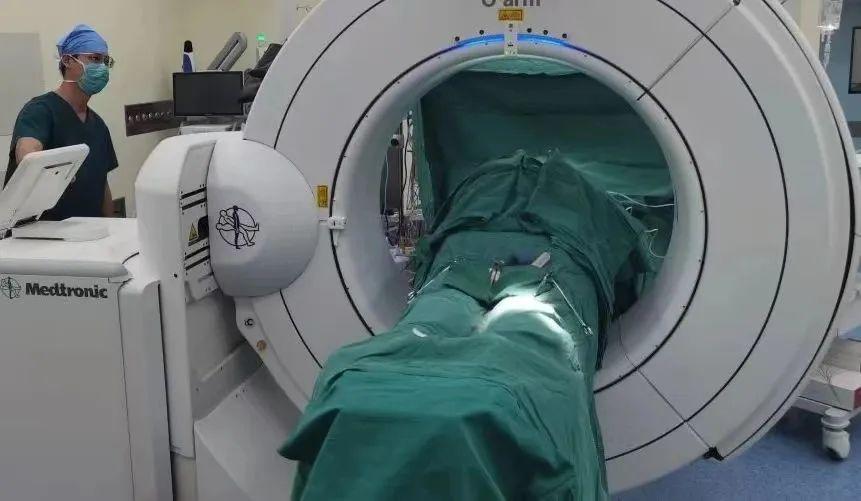
Recently, the team from the Department of Orthopedics and Oncology at Peking University People's Hospital completed the first domestic surgery for sacral tumor resection in the sagittal plane under the guidance of the "O" arm navigation.
This navigation technology achieves precise tumor localization during surgery, preserving the contralateral sacral nerve while completely removing the sacral tumor for a 35 year old young patient, maximizing the protection of surrounding normal bone, important blood vessels, and organs, significantly reducing intraoperative bleeding, and greatly shortening the patient's postoperative recovery time.
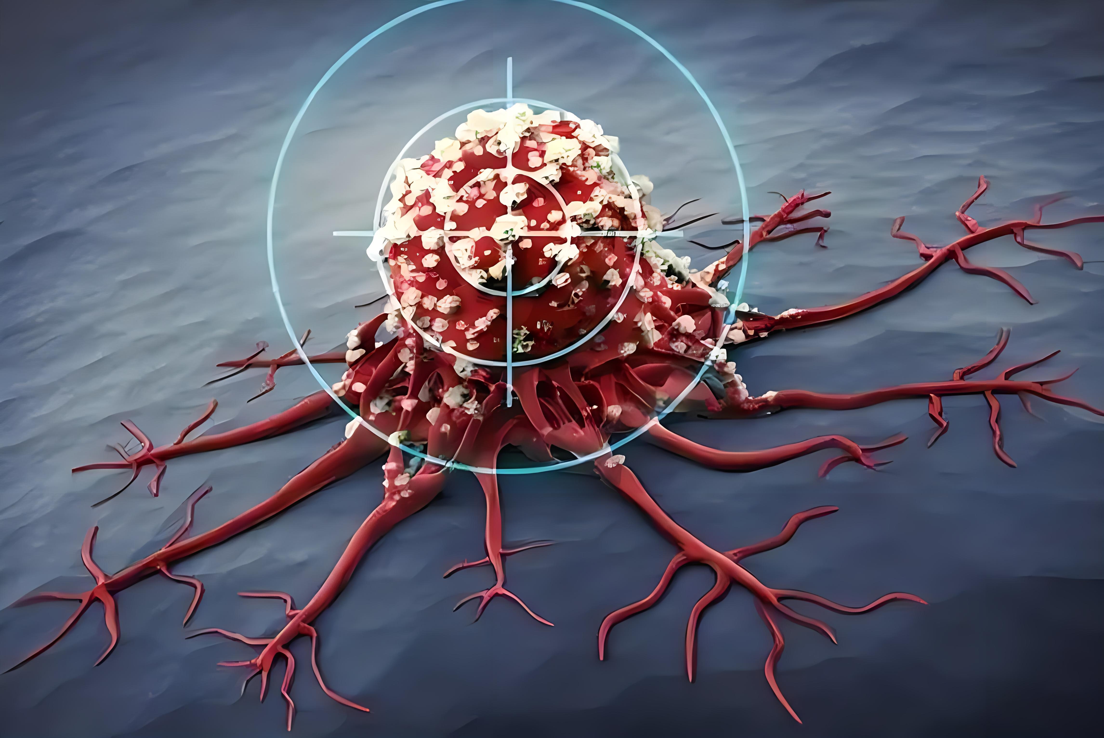
The pain in the tail bone is actually due to malignant tumors
Xiao Wang (pseudonym) is a 35 year old patient who started experiencing sacrococcygeal pain 5 months ago. Initially, he didn't take it seriously, but the symptoms worsened and normal walking became difficult.
Go to the local hospital for treatment, the imaging examination indicates sacral bone lesions, the biopsy pathology suggests malignant tumors with epithelioid differentiation, and the PET-CT examination suggests single bone lesions.
Sacral malignant tumors are a more serious disease, and their epithelial differentiation makes them even rarer. I didn't expect such a serious illness to be hidden behind the pain. Recommended by the local hospital, the young man and his family immediately rushed to the Department of Orthopedics and Oncology at Peking University People's Hospital in search of hope, placing their life-saving straw here.
One of the most difficult surgeries in sacral tumor resection surgery
Surgical treatment is one of the main treatment methods for sacral malignant tumors. In the field of bone tumor treatment, the surgical resection of sacral tumors, spinal tumors, and pelvic tumors is collectively known as the three most technically challenging surgeries.
Due to the deep position of the sacrum and the proximity of many important blood vessels, nerves, and tissue structures, the surgical risk is relatively high. But if left unchecked, it may spread and metastasize, leading to serious complications such as urinary and fecal dysfunction, sexual dysfunction, sacrococcygeal pain, etc. But Professor Tang Xiaodong, the director of the Department of Bone Oncology, still decided to give this young life a fight.
This decision is inseparable from years of accumulated experience. After thirty years of unremitting efforts and technological inheritance, the Orthopedic Oncology Department of Peking University People's Hospital has accumulated profound experience in the treatment of sacral tumors, transforming this high-risk surgery into a safe and controllable routine operation.
We provide treatment for numerous sacral tumor patients every year, becoming the bone tumor treatment center with the highest number of such surgeries completed globally.
While achieving significant achievements, Professor Tang Xiaodong took over the baton from his predecessors and led the team to continuously explore, aiming to improve the thoroughness of tumor resection while minimizing harm to patients and preserving more functions.
"O" arm navigation assists in precise tumor resection
The doctor found that Xiao Wang's tumor had already affected the left sacrum and was accompanied by a soft tissue mass. Fortunately, Xiao Wang had a single bone lesion, and the tumor was limited to one bone and did not spread to other areas. Medical staff have jointly engaged in a race with tumor cells.
Before surgery, fusion imaging is performed on the MRI and CT images of the tumor, allowing the surgeon to have a 3D understanding of the tumor's extent. Based on the image information, the doctor formulates the resection path and range, and determines a detailed preoperative plan.
3D fusion imaging suggests tumor involvement of the left sacrum, accompanied by soft tissue mass
During surgery, doctors need to rely on rich experience and professional skills to preserve the function of nerves and blood vessels as much as possible, while thoroughly removing tumor tissue to reduce the risk of recurrence and metastasis. But it is difficult to achieve precise and thorough tumor resection with the naked eye.
The "O" arm navigation system is an advanced orthopedic surgical auxiliary device that can accurately locate tumors. This system consists of an "O" arm intraoperative imaging and surgical navigation system, playing an important role in orthopedic surgery. It provides real-time three-dimensional images for surgeons, allowing them to have a broader perspective and helping them avoid important blood vessels and nerves in the surrounding area. It accurately locates the lesion site and range, ensuring complete tumor resection and maximum preservation of normal bone, greatly improving surgical safety.
After precise resection of the tumor, reconstruction was performed using a 3D printed composite half sacral prosthesis independently developed by the Department of Orthopedics and Oncology. And under the guidance of the O-arm navigation system, the prosthesis and screws are precisely placed to further improve the reconstruction effect of the patient. In addition, the system can also shorten surgical time and accelerate patient recovery.
The first domestic case of precise sagittal resection of sacral tumors under "O" arm navigation
This surgery not only marks progress in the field of bone tumor treatment, but also highlights our ability to ensure thorough tumor resection while greatly protecting healthy tissues, which is of critical significance for postoperative functional improvement of patients.
Director Tang Xiaodong stated that the successful application of this technology not only brings new hope to patients with bone tumors, but also accumulates valuable experience for precise treatment of bone tumors, opening up a path towards a healthier future. The team plans to expand the application of O-arm navigation technology from spinal and pelvic surgery to limb tumor resection, further optimizing limb and joint preservation surgery, and bringing greater benefits to patients.
Contributed by Zang Jie and Tang Xiaodong from the Department of Bone Oncology
Editor | Zhong Yanyu, Zhang He
Statement | Reproduction source: Peking University People's Hospital“


COMMENT